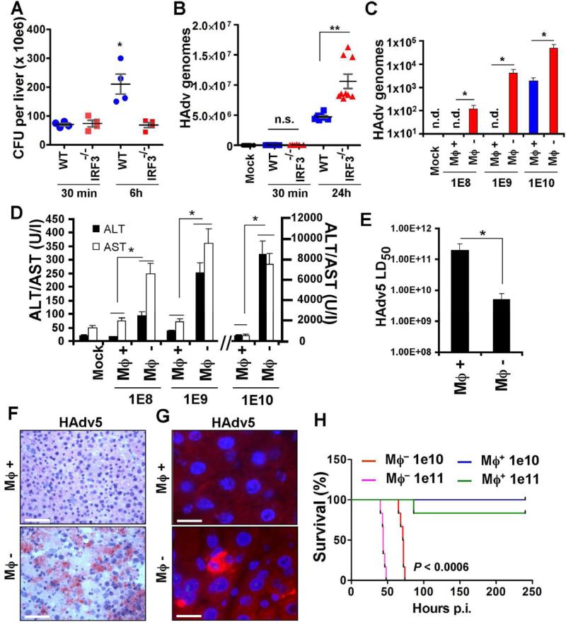
Figure 4. IRF3-dependent “defensive suicide” is a protective macrophage effector mechanism against disseminated virus infection.
Pathogen burden in the livers of WT and Irf3-/- mice infected with L.monocytogenes (A) and wild type HAdv5 (B) analyzed at indicated times. Representative data from two experiments are shown. The data in (A) was analyzed by 1-way ANOVA, * P = 0.0014, and in (B) by 1-way ANOVA with post-hoc Kruskal-Wallis test. ** P < 0.001. (C) The amounts of the virus genomic DNA in the livers of wild type mice depleted of tissue macrophages with clodronate liposomes (Mϕ -) or mice containing tissue macrophages (Mϕ +; mice were injected with liposomes containing saline only) 48 hours after challenge with HAdv5 at indicated doses (in virus particles per mouse). The amounts of viral genomes per 10 ng of total liver DNA are shown. N = 6. * P < 0.01. (D) The amounts of liver enzymes ALT and AST in plasma Mϕ- and Mϕ+ mice 48 hours after challenge with HAdv5 at indicated doses. In Mock settings, ALT and AST levels were similar in Mϕ+ and Mϕ- mice prior to virus challenge. N = 6. * - P < 0.01. (E) Reduction of LD50 of HAdv5 for mice after depletion of tissue macrophages with clodronate liposomes (Mϕ-), compared to mice containing tissue macrophages (Mϕ+). N = 9. * P < 0.05. (F) Hematoxyllin and eosin staining of liver sections from Mϕ+ and Mϕ- mice 48 hours after infection with 1010 virus particles of HAdv5. Scale bar is 100 μm. (G) Sections of liver as in (F) after staining with anti-HAdv5 hexon Ab (red). Cell nuclei were couterstained with DAPI (blue). Scale bar is 10 μm. (H) Survival of Mϕ- and Mϕ+ mice after challenge with indicated doses of HAd5. N = 8. Statistical significance of P < 0.0006 is indicated for groups of Mϕ- mice when compared to Mϕ+ mice using log-rank test.

