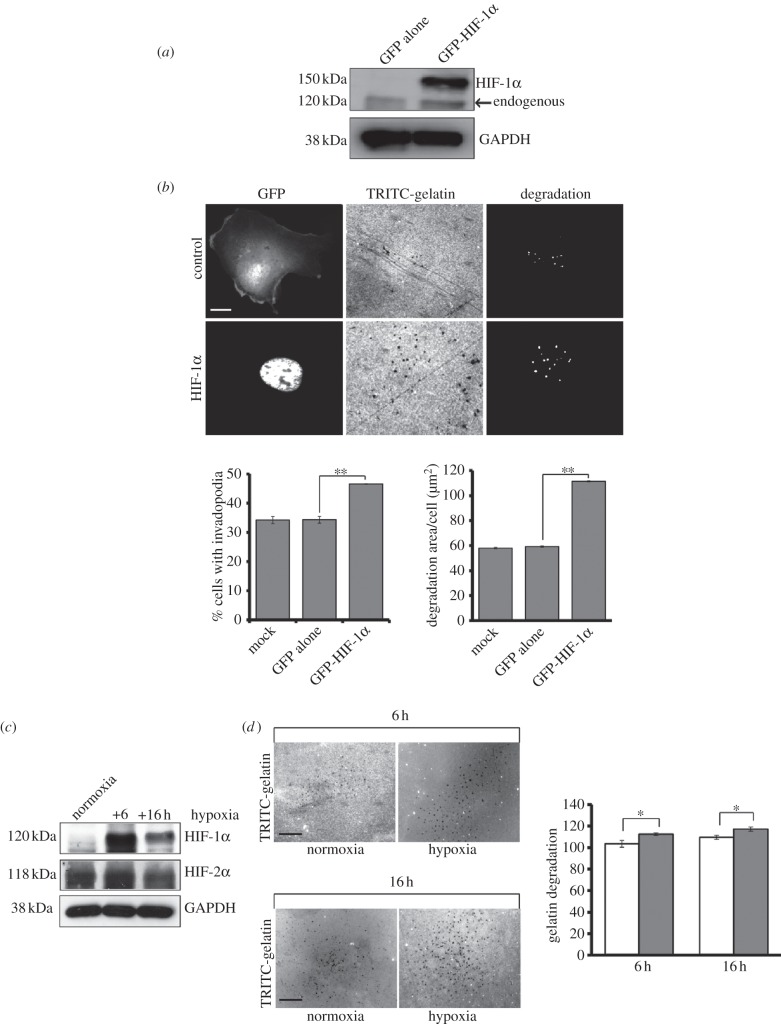
Figure 2.
Specific HIF-1α overexpression induces invadopodia formation. (a) Lysates from GFP-HIF-1α-transfected cells were probed for HIF-1α expression and GAPDH. (b) GFP-HIF-1α-transfected cells were seeded on gelatin coverslips, fixed and stained for F-actin. Cells were scored for the presence of GFP-HIF-1α expression in the nucleus and corresponding actin puncta that colocalize with area of degradation on the gelatin. A gelatin degradation assay was performed using ImageJ software. (c) Cells were incubated in normoxia or hypoxia incubators for 6 and 16 h. Cell lysates were probed for levels of HIF-1α and re-probed for HIF-2α. GAPDH was used as a loading control. (d) Cells were seeded on gelatin-coated coverslips and incubated in normoxia (open bars) or hypoxia (filled bars) incubator for 6/16 h. The area of degradation on the gelatin was measured using ImageJ software. The results shown are mean grey value ± s.e.m. of 30 fields of view from each experimental condition over three separate experiments. Statistical significance was calculated using Student's t-test; *p < 0.05, **p < 0.005, scale bar = 10 µm.

