Figure 4.
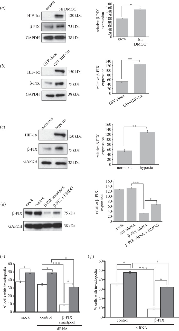
β-PIX is required for invadopodia formation. (a) DMOG stimulated, (b) GFP-HIF-1α overexpressing cells and (c) hypoxic cell lysates were probed for expression of HIF-1α, β-PIX and GAPDH as a loading control. (d) MDA-MB-231cells were transfected with mock, control and β-PIX siRNAs for 24 h, ±DMOG for 6 h. Cell lysates were probed for levels of β-PIX and GAPDH as a loading control. (e) MDA-MB-231 cells transfected with mock, control and β-PIX siRNAs for 24 h, ±DMOG (+, filled bars; –, open bars) for 6 h were seeded on gelatin-coated coverslips. Cells were scored as figure 1c. (f) Cells expressing GFP alone (open bars) or GFP-HIF-1α (filled bars) were transfected with β-PIX siRNAs for 24 h then seeded on gelatin-coated coverslips. Cells were scored as figure 1c. In all cases, relative expression of β-PIX was measured using densitometric analysis over three separate experiments. In all cases, the results shown are mean±s.e.m. of 30 cells from each experimental condition over three separate experiments. Statistical significance was calculated using Student's t-test; *p < 0.05, ***p < 0.0005.

