Figure 1.
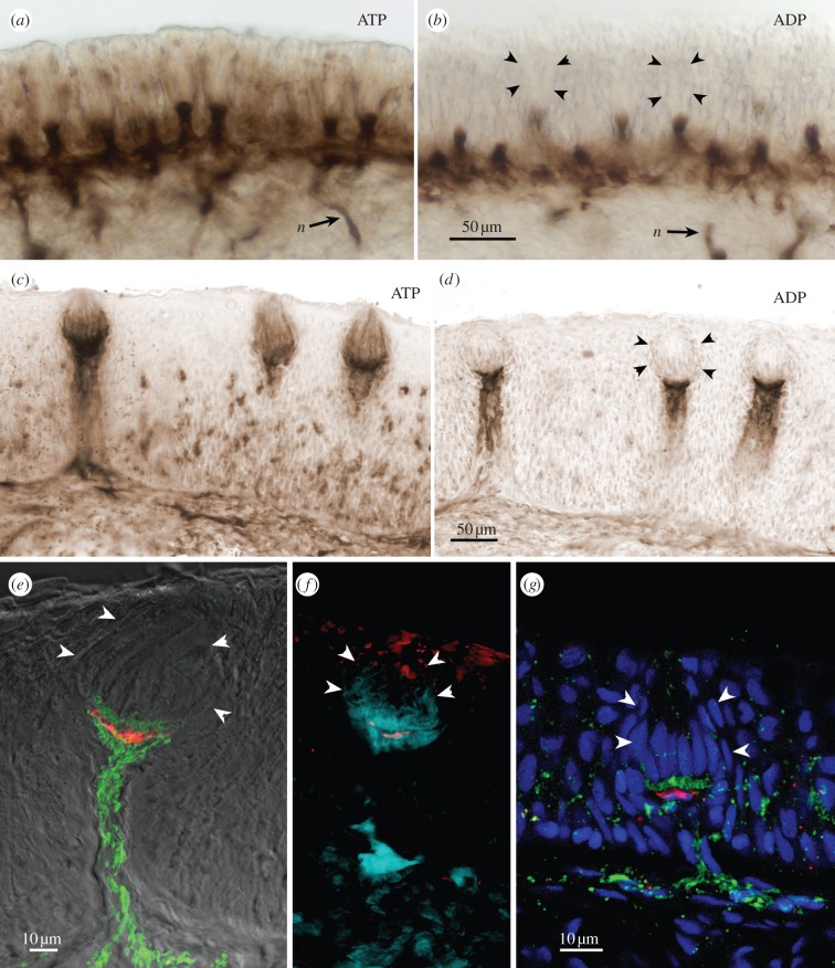
(a–f) Ecto-ATPase and non-specific nucleotidase (ADP) staining in taste buds from goldfish, C. auratus. (a,b) Sections through the palatal organ showing (a) specific ecto-ATPase activity, and (b) non-specific nucleotidase activity. (c,d) Sections through the lip showing (c) specific ecto-ATPase activity, and (d) non-specific nucleotidase activity. In (a–d), Ecto-ATPase staining is evident in the elongate cells of the taste bud as well as in the basal portion of the taste bud proper. Non-specific staining with ADP as substrate (b,d) shows reaction product surrounding the vertical nerve bundles and extending to a pedestal below the taste bud. (e,f) Higher magnification views of single taste buds showing serotonergic immunoreactivity (red) of the (e) Merkel-like basal cells in lip and (f) palatal organ. The ecto-ATPase activity is shown in pseudocolour: green in (e) and aqua in (f). The pseudocolour image is produced by inverting a brightfield image and placing into a colour channel of the composite image from tissue first reacted for ecto-ATPase and then immunoreacted for serotonin. Note that the ecto-ATPase staining appears both above and below the Merkel-like basal cell. (g) Longitudinal section through the taste bud from the lip of a P2X3a-GFP zebrafish showing that nerve fibres expressing purinergic receptors (green) form a plexus mostly above the Merkel-like basal cell immunoreacted for serotonin (red). In all panels, arrowheads indicate the edge of the taste bud.

