Figure 3.
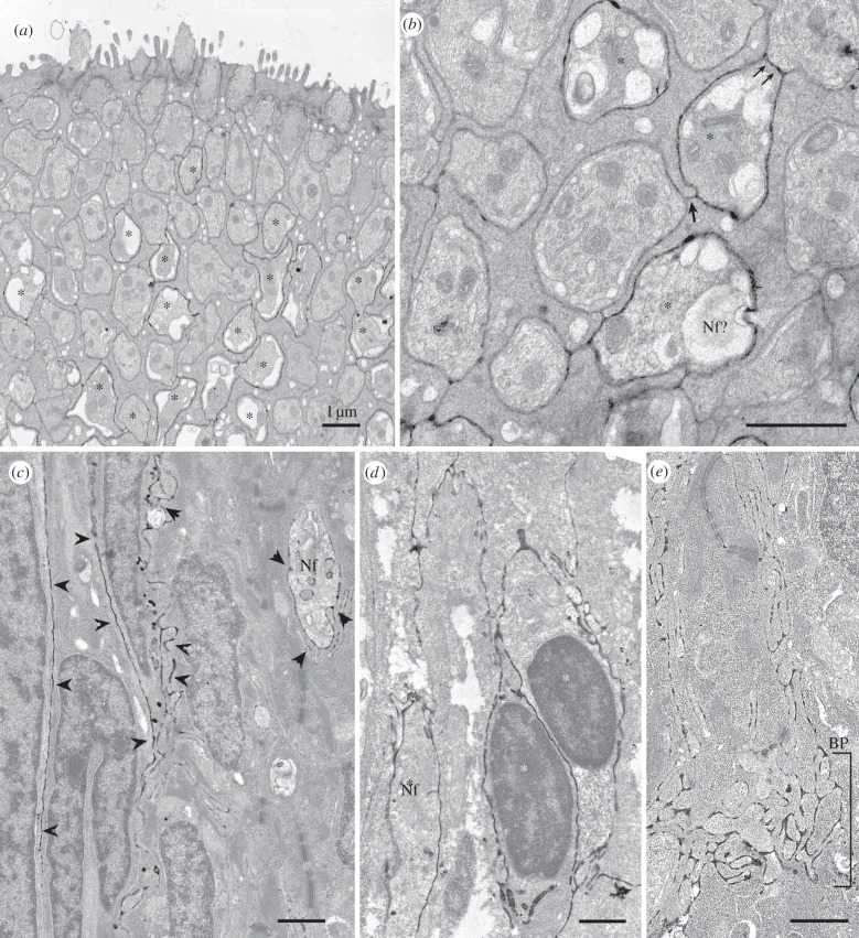
Electron micrographs of ecto-ATPase staining of taste buds from (a,b) the catfish, I. punctatus and (c–e) goldfish. (a,b) Cross sections through the apical region of a barbel taste bud showing that many but not all light cells are surrounded by reaction product. (a) Low magnification image showing numerous light cells (asterisks) surrounded by label at a depth of about 5 µm from the taste pore but largely devoid of label higher in the taste bud. As described by Reutter (reviewed in [27]), the light cells terminate apically in a large single microvillous process. Dark cells surround the light cells and end apically with a small tuft of microvilli. (b) Higher magnification view of another section from the same specimen showing details of the distribution of the reaction product. Several light cells (asterisks) are surrounded by reaction product. In one case (double arrow), a reactive light cell (asterisk) directly contacts an unlabelled light cell and the ecto-ATPase reaction product continues across this line of contact, strongly suggesting that the plasma membrane of the labelled light cell houses the ecto-ATPase enzyme. Conversely, at the junctional face of two non-reactive dark cells (single arrow), we see slight evidence of reaction product probably due to drift of the ecto-ATPase reaction product from the adjacent labelled light cells. This shows the limitations of ultrastructural analysis with this histochemical technique. (c–e) Longitudinal sections through the taste buds of a goldfish. (c) Section through the taste bud from the lip showing elongate cells surrounded by reaction product (arrowheads). In addition, a profile of a nerve fibre (Nf) is similarly surrounded by reaction product. (d) Palatal taste bud also revealing elongate cells (asterisks) surrounded by reaction product. (e) Section through the basal region of a labial taste bud showing strong reaction product surrounding many nerve fibres within the BP. The nerve fibres at this level are not surrounded by any glial elements and so the reaction product is most probably produced by ectoenzymes on the membranes of the nerve fibres themselves. This is different than the situation in rodent taste buds where the ectoenzyme is mostly associated with the membranes of the type I taste cells [13].

