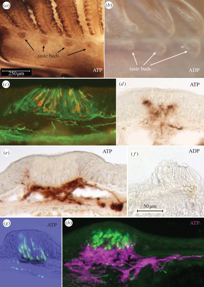
Figure 5.
Taste buds in lamprey show specific ecto-ATPase reactivity. (a,b) Whole mount staining of the branchial apparatus of the lamprey showing (a) Ecto-ATPase staining, and (b) paucity of non-specific nucleotidase staining. (c) Staining of the taste bud for acetylated tubulin (green) and serotonin (red) shows that all cells of the taste bud are elongate cells; no basal Merkel-like cells exist (also see [37]). (d,e,f) Sections reveal that the specific ecto-ATPase activity (d,e) occurs along the basal portions of the taste bud with some lateral staining extending slightly upwards at the margins of the taste bud. With the ADP substrate (f) little staining is seen. (g) Section through a taste bud showing ecto-ATPase staining (black) and serotonin immunoreactivity (green). The basal end of the serotonin-positive cells inserts into the area occupied by ecto-ATPase staining. (h) An oblique section through a taste bud showing serotonin staining (green) and pseudocoloured ecto-ATPase activity (magenta). The serotonergic cells extend a basal process apparently touching and embedded within the ecto-ATPase positive regions. Fibrillar ecto-ATPase staining is present under the basal lamina in association with nerve fibres. Some serotonergic fibres also are evident, but are not associated with the ecto-ATPase staining.

