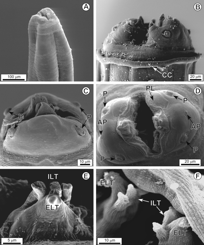
Figure 2.
Physaloptera ngoci. A, cephalic extremity, general view. B, cephalic extremity, lateral view, cephalic collarette (CC). C, cephalic extremity with detail of papillae (P), lateral view. D, cephalic extremity, apical view. AP, amphids; PL, porus-like region; P, papillae. E, F, detail of externolateral tooth (ELT) and tripartite internolateral tooth (ILT).

