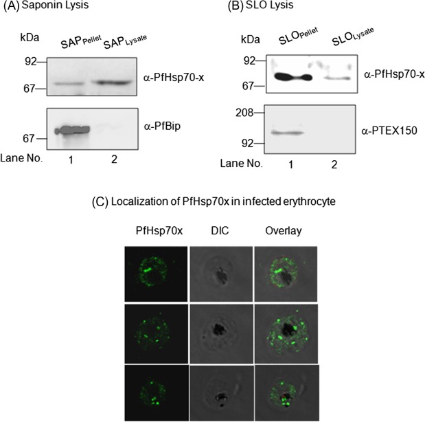
Figure 5.
Sub-cellular localization of PfHsp70-x in infected erythrocyte: Infected erythrocytes were lysed with saponin (A) and SLO (B) and the fractions obtained were analysed by immunoblotting with PfHsp70-x antiserum (top). To ensure that compartmental integrity was maintained during saponin and SLO-based fractionation, the fractions were immunoblotted with PfBip and PTEX150 respectively (below). PfHsp70-x gets exported outside the parasite with major population being present in the PV and some in the erythrocyte compartment. (C) IFA analysis of infected erythrocytes with PfHsp70-x antiserum reveals its presence throughout the infected erythrocyte.

