
Figure 2.
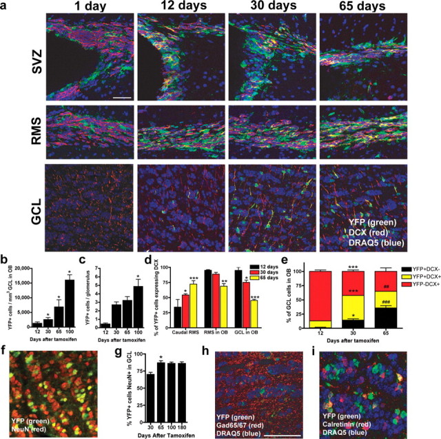
Neurogenesis in the SVZ/RMS/OB following TAM. a, Time course for the appearance of YFP+ cells in the SVZ, RMS, and OB GCL 1, 12, 30, and 65 d after TAM. Recombined cells (YFP+, green) are present 1 d after TAM within the SVZ, whereas fewer YFP+ cells are present in the RMS and virtually no recombined cells are present within the GCL. With increasing time after TAM, there are many recombined cells in each of these areas with a variable proportion of these cells being neuroblasts as demonstrated by coexpression of YFP and DCX (red). b, c, YFP+ cells in the OB GCL (a) and OB GL (b) significantly increased up to 100 d (*p < 0.05 vs 12 d). d, The proportion of YFP+ cells that are migrating neuroblasts (DCX+) increased in the caudal RMS but decreased in the RMS in OB and GCL in OB (*p < 0.05, **p < 0.01, and ***p < 0.005 vs 12 d). e, The proportion of YFP+ cells (black and yellow bars) that are not migrating neuroblasts (black bars) increased in the OB GCL (*p < 0.05 and ****p < 0.005 vs 12 d; ##p < 0.01 and ###p < 0.005 vs 30 d). f–i, Thirty days after TAM, most recombined cells (f, g) in the GCL were NeuN+ (*p < 0.05 vs 30 d) as well as GABAergic interneurons (h, i), as assessed via colabeling with YFP and GAD65/67 (h; counterstain DRAQ5) and CR (i). Scale bar: f, h, i, 50 μm. Error bars indicate SEM.