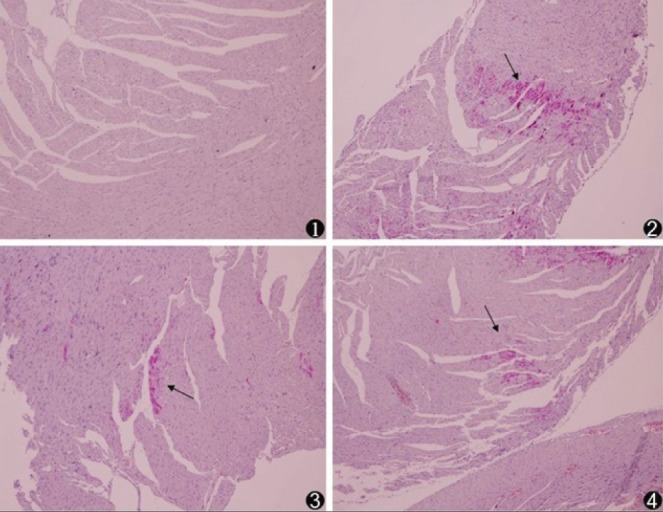
Figure 2).

Histopathology of myocardial microinfarcts (original magnification ×100). Hematoxylin-basic fuchsin-picric acid staining of tissue samples revealed no infarct in the sham-operated (control, 1) animals. Microinfarcts (arrows) in the tissue samples from coronary microembolization (2), metoprolol (3) and Z-LEHD-FMK (4) groups. Normal myocardial cytoplasm is stained yellow, the nuclei are stained blue and the ischemic myocardium is stained red
