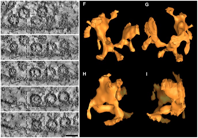
Figure 3. Shape of the luminal assembly of macromolecules in the principal vesicle.
(A–E) Selected 1 nm thick virtual slices from a series made through one of the primary reconstructions. The section from which the reconstruction was made was cut in the active zone’s median plane; the virtual slices are shown in the same plane. Four vesicles are docked in a row on the presynaptic membrane (PM). Undocked vesicles are nearby. All of the vesicles contain luminal assemblies of macromolecules. The box in each virtual slice outlines the so-called principal vesicle. (F–I) 3D surface model of the principal vesicle’s luminal assembly shown in different degrees of rotation. In (F) the assembly is viewed from the median plane of the AZM with the portion facing the presynaptic membrane downward. In (G) the assembly is rotated 180 degrees around the vertical axis of its orientation in (F). In (H) and (I) the assembly is rotated 90 degrees to the right and left, respectively, around the vertical axis of the orientation in (F). The assembly has a bilateral arrangement of four irregular arms, which radiate from below and behind the center of the vesicle. Nubs of varying lengths arise from the arms. Scale bar (A–E) = 50 nm.

