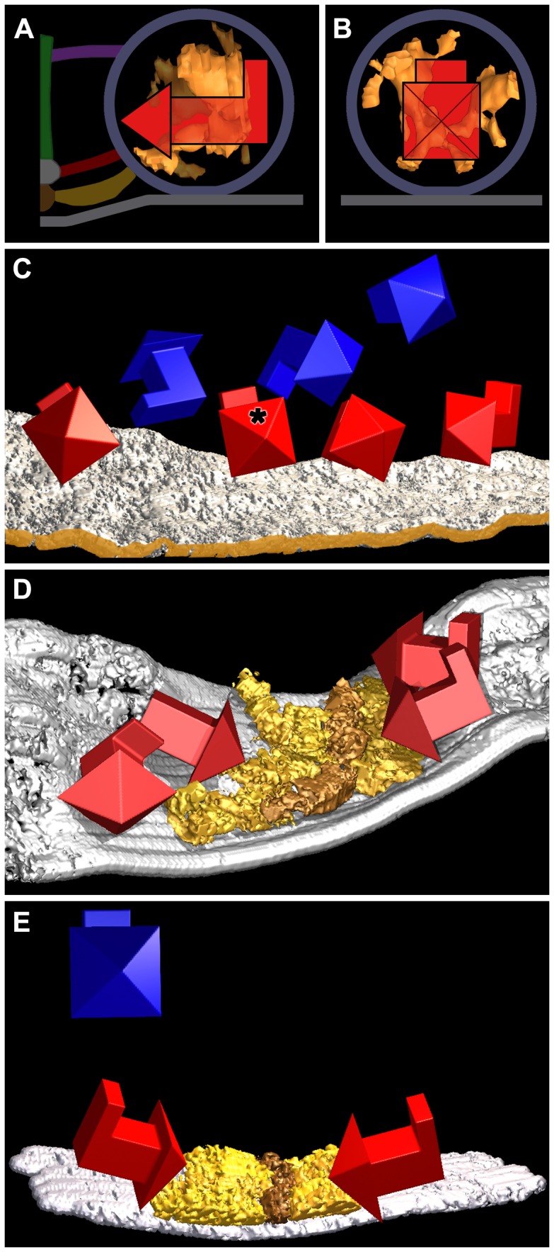
Figure 5. Orientation of the shape of the luminal assemblies.

In (A), the surface model of the luminal assembly of the principal vesicle is viewed in the transverse plane of its active zone, while in (B) it is viewed from the median plane of the active zone. The 3D arrow superimposed on the surface model in both (A) and (B) is oriented so its head points to the median plane of the AZM, its shaft is orthogonal to the median plane and parallel to the presynaptic membrane and its tail is vertical to the presynaptic membrane. The vesicle membrane, presynaptic membrane, and AZM macromolecules are schematized for reference. (C) A surface model of the presynaptic membrane in a primary reconstruction, shown in the virtual slices in Figure 3A–E that include the principal vesicle, three other docked vesicles and three undocked vesicles. The edge of the membrane nearest the median plane of the AZM is brown-gold. The 3D arrows show the orientation of the shape of the luminal assembly in the docked (red) and undocked (blue) vesicles relative to that of the principal docked vesicle (asterisk) with respect to the median plane of the active zone and to the presynaptic membrane as shown in (A) and (B). The shape of the assembly in all of the docked vesicles has the same orientation (±30 degrees) as that of the principal vesicle, while the shape of the assembly in undocked vesicles does not share a common orientation. (D) Surface models of the presynaptic membrane and superficial layer of the AZM from the other primary reconstruction. The superficial layer of the AZM, which contains ribs (gold) and beams (brown-gold), is viewed from near the active zone’s transverse plane. There is a slight angular change midway along the AZM’s long axis [11]. The 3D arrows show the orientation of the shape of the luminal assembly in four docked vesicles relative to that of the primary docked vesicle in (A–C) with respect to the median plane of the active zone and presynaptic membrane. The shape of the assembly for three of the docked vesicles had the same orientation (±30 degrees) with respect to the median plane of the active zone and to the presynaptic membrane as the docked vesicles in (C). While the shape of the assembly for the fourth vesicle (arrowhead and shaft lacking a tail) was similarly oriented with respect to the median plane of the active zone, there was not sufficient information to determine its orientation with respect to the presynaptic membrane. (E) Surface models of the presynaptic membrane and superficial layer of the AZM from another reconstruction. The ribs and beams of the AZM are viewed from near the active zone’s transverse plane. The 3D arrows show the orientation of the shape of the luminal assembly in two docked vesicles (red) and one undocked vesicle (blue) relative to that of the primary docked vesicle in (A–C) with respect to the median plane of its active zone and presynaptic membrane. The shape of the assembly for the two docked vesicles had the same orientation (±30 degrees) with respect to the median plane of the active zone and to the presynaptic membrane as the docked vesicles in (C) and (D), while the shape of the assembly in the undocked vesicle did not.
