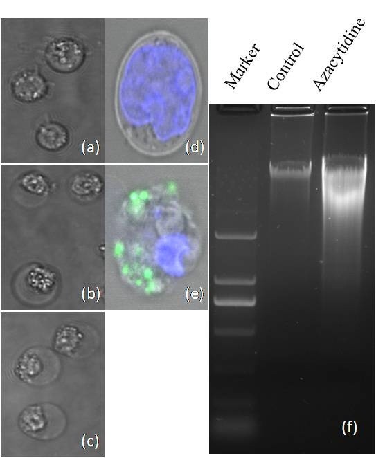
Figure 2.

Morphologic images of myeloma cells. (a) untreated cells; (b) 100 μM azacytidine-treated cells for 24 h; and (c) H2O2-treated U266 cells; All images were captured by Olympus IX2-UCB 60× inverted microscopy; (d-e) detection of ROS in untreated and azacytidine-treated U266 cells using the Image-iT LIVE Reactive Oxygen Species (ROS) Kit. Cells were labeled with carboxy-H2DCFDA, which exhibited green fluorescence when reacted with ROS, and nuclei were stained with blue-fluorescent Hoechst 33342. (d) untreated U266 cells; and (e) azacytidine-treated cells; and (f) gel electrophoresis of DNA from untreated and azacytidine-treated myeloma cells.
