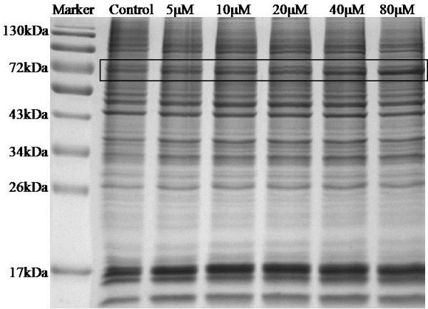
Figure 3.

The 1D SDS-PAGE gel image of untreated and 24 h, azacytidine-treated myeloma cells. Lane 1, molecular weight markers; Lane 2, proteins from untreated cells; Lane 3, proteins from 5 μM azacytidine-treated cells; Lane 4, proteins from 10 μM azacytidine-treated cells; Lane 5, proteins from 20 μM azacytidine-treated cells; Lane 6, proteins from 40 μM azacytidine-treated cells; and Lane 7, proteins from 80 μM azacytidine-treated cells;. The band with the most differentially expressed proteins was marked with a square.
