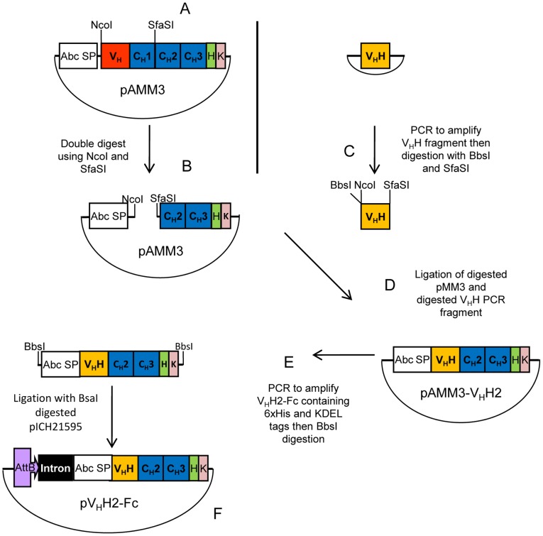
Figure 1. Schematic representation of the VHH2-Fc antibody cloning strategy.
(A) pAMM3 was created by adding a SfaSI restriction enzyme site to pMM3 using PCR mutagenesis. (B) pAMM3 was digested with NcoI and SfaSI to remove VH and CH1 domains and retaining the Fc of IgG1. (C) VHH C2 was PCR amplified and then digested with BbsI and SfaSI. (D) Digested pAMM3 and VHH C2 were ligated together thereby fusing the VHH with the Fc. (E) VHH2-Fc coding sequence was PCR amplified from pAMM3-VHH2, then digested with BbsI. (F) Digested VHH2-Fc fragments were ligated with BsaI-digested pICH21595 generating pVHH2-Fc construct. Restriction enzymes sites: NcoI, SfaSI, BbsI and BsaI.

