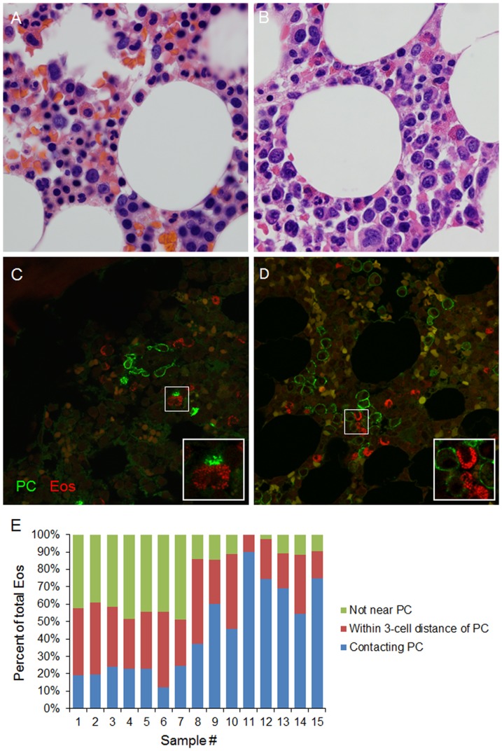
Figure 1. Eosinophils and PCs are found in close proximity in the human bone marrow.
BM biopsies were obtained from normal donors (A, C) and MM patients (B, D) and stained with H&E (A, B) or using immunofluorescence (C, D) for selective visualization of PCs and Eos. In immunofluorescence staining, PCs were stained with anti-CD138 mAb (green) and Eos were stained with chromotrope 2R (red). Autofluorescent red blood cells are shown in yellow in these overlaid images. Images are representative of 5 normal donor and 10 MM patient BM biopsies. (E) Quantitation of Eos across 6 random fields from each immunofluorescence-stained sample dividing Eos into 3 categories: 1) Eos in direct contact with PCs; 2) Eos within a 3-cell distance of PCs; and 3) Eos more than a 3-cell distance away from the closest PC. Samples #1-5 are BM biopsies from normal donors. Samples #6–8 are from patients with MGUS. Samples #10–12 are from patients with SMM. Samples #9 and 13–15 are from patients with MM. See Table S1 for more detail.

