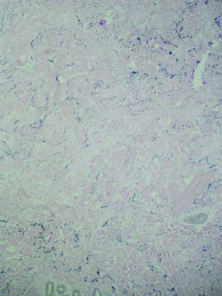
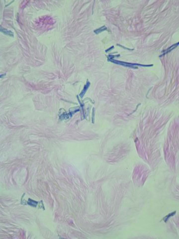
Figures 2A and 2B.


Microscopic examination—Intermediate (A) and higher (B) magnification—of the linear, raised, horizontal, lumbar bands show a localized lysis of the elastic fibers in the mid dermis: There is a paucity of fragmented elastic fibers in the upper portion of the reticular dermis. In contrast, normal-appearing elastic fibers are present in both papillary dermis and the deep reticular dermis (Verhoeff-van Gieson: A=X10; C=X20).
