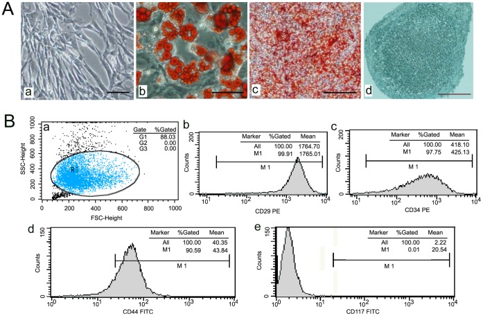
Figure 1. The identification of BM-MSCs.
(A) The morphology of MSCs from male mice was observed under light microscopy. Bar, 100 um (a). mMSCs were induced to differentiate into adipocytes. Bar, 75 um (b), osteoblasts. Bar 400 um (c), or chondrocytes Bar, 75 um (d) using an in vitro differentiation assay. (B) The surface marker of MSCs were detected flow cytometry analysis(a), positive for CD29(b), CD34(c) and CD44(d), but negative for CD117 (e).

