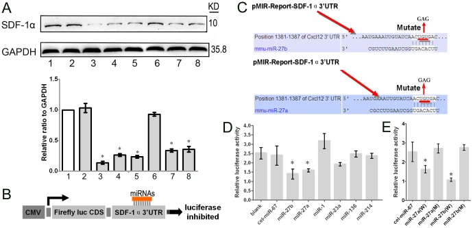
Figure 5. The effect of the miRNAs on the level of SDF-1α and luciferase of the 3′UTR.
(A) The expression of SDF-1α protein in MSCs after 96-hour infection with lentiviral vectors expressing the given miRNA, detected by western blot. Lane 1, MSCs; lane 2, MSCs/cel-miR-67; lane 3, MSCs/miR-27b; lane 4, MSCs/miR-27a; lane 5, MSCs/miR-1; lane 6, MSCs/miR-23a; lane 7, MSCs/miR-136; lane 8, MSCs/miR-214. The relative expression ratio to GAPDH was summarized in the below panel. (B) The amplified wild-type SDF-1α 3′UTR was cloned into the dual-luciferase miRNA target expression vector, and potential binding sites for miRNAs in the 3′UTR of SDF-1α were predicted by computational analysis. (C) To test for potential interactions with miRNAs, the SDF-1α 3′UTR and the given miRNA were co-transfected into HEK293 cells. The activities of Firefly and Renilla luciferase were assayed 24 h post-transfection (n = 3, *p<0.05; Mann-Whitney test). The error bars represent S.D. (D) The predicted miR-27a and miR-27b binding sites in SDF-1α were mutated by site-directed mutagenesis of the miRNA seed region. (E) The Firefly and Renilla luciferase activities were analyzed in HEK293 cells 24 hours after co-transfection with the given miRNA and the mutated SDF-1α 3′UTR. (n = 3, *p<0.05; Mann-Whitney test). The error bars represent S.D.

