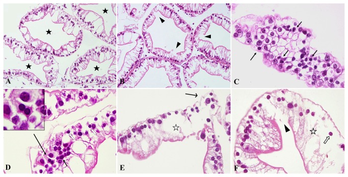
Figure 1. Morphological injury induced by Cd in S . henanense hepatopancreas with paraffin sections and HE staining (n = 6).
(A) Normal tissue in the control group, ×200. The hepatopancreas glands were circular or elliptical, and the inner surface was irregular but continuous. (B) A few cells were damaged and the continuation of the inner surface was broken in the 3.56-mg/L Cd group, ×200. (C) Transverse section of the hepatopancreatic gland in the 3.56-mg/L Cd group, ×600. Cells arrange like the honeycomb. Classical apoptosis bodies (arrow) with chromatin aggregation and more acidophilia of cytoplasm were smaller than the surrounding ones. (D) Apoptosis was easily identifiable in the 7.12-mg/L Cd group, ×600. Classical apoptotic bodies are shown in the upper left corner. (E) Severely cellular swelling and damage were present in the 28.49-mg/L Cd group, ×400. (F) Broken epithelia, grave cell swelling and necroptosis were present in the 56.98-mg/L Cd group, ×600 (★: hepatopancreas glandular cavity; ◄: broken epithelia; ➞: apoptosis; ☆: severely cellular swelling; ➔: deciduous cells; ⇧: necroptosis).

