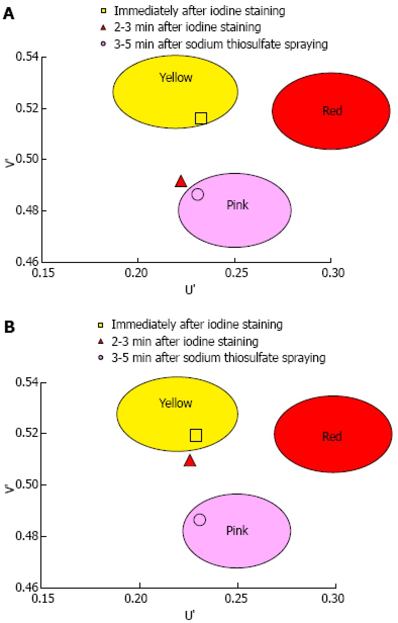
Figure 5.

Mean U’ and V’ values of the mucosa in the early, late and final phases were plotted on a color diagram. A: Pink-color sign positive mucosa had a lower mean V’ value in the late phase (pinkish color) than in the early phase (yellowish color), suggesting that the pink-color sign positive mucosa underwent a color change from yellow to pink. The mucosa had similar mean U’ and V’ values in the late and final phases, suggesting that the color of pink-color sign positive mucosa in the late stage was similar to the color of mucosa after complete fading of iodine staining; B: Pink-color sign negative mucosa had similar mean U’ and V’ values in the early and late phases (yellowish color), suggesting that pink-color sign negative mucosa did not change in color during this time period.
