
Figure 1.
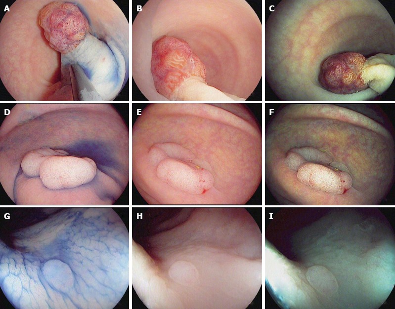
Characterization of colorectal polyps using chromoendoscopy with indigo carmine 0.4% (A, D and G) and high-definition i-scan, surface enhancement (B, E and H)/tone enhancement (C, F and I), the i-scan classification for endoscopic diagnosis. A-C: 20 mm sized pedunculated polyp (Paris Ip). Image enhanced endoscopy shows reddish color, prominent vessels and a type IV pit pattern of the epithelial surface. Histopathology showed a tubulovillous adenoma with low-grade dysplasia; D-F: 40 mm sized non-polypoid (Paris IIa) lesion. Image enhanced endoscopy shows reddish color, dilated, irregular vessels and a type IV pit pattern of the epithelial surface. Histopathology showed a tubulovillous adenoma with high-grade dysplasia; G-I: 3 mm sized non-polypoid (Paris IIa) lesion. Image enhanced endoscopy shows pale color, isolated, lacy vessels and a type II pit pattern. Histopathology showed a hyperplastic polyp.