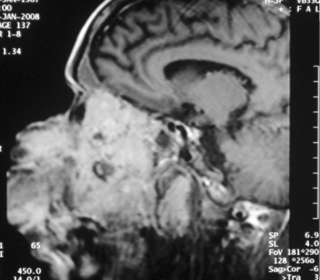
Fig. 2.

MRI showed homogenous soft tissue mass extending from subcutaneous tissue on left side of face to left infratemporal fossa, inferiorly oral cavity with destruction of hard palate and superiorly involving left orbit with destruction of medial and inferior wall with ill defined fat planes. It was hypointense on T1 and hyperintense on T2 and showed moderate contrast enhancement
