Figure 3.
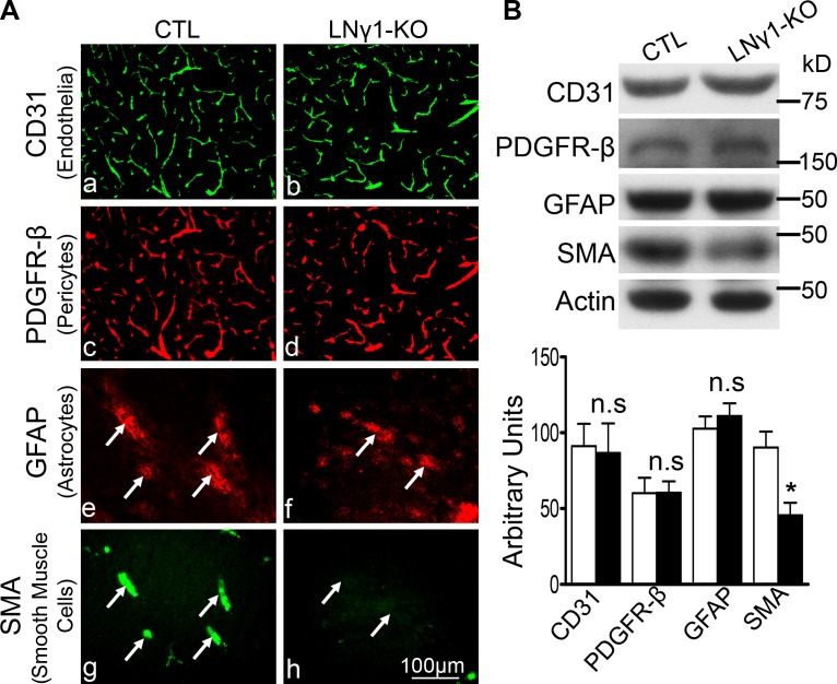
Cerebral vascular wall changes in the striatum of LNγ1-KO mice. (A) Immunohistochemistry analysis revealed expressions of CD31 (endothelial cell marker; Aa and Ab), PDGFR-β (pericyte marker; Ac and Ad), and GFAP (astrocyte marker; Ae and Af) were similar between control and LNγ1-KO mice. However, expression of SMA (smooth muscle cell marker) was decreased in the striata of LNγ1-KO compared with control mice (Ag and Ah). GFAP expression was associated with SMA in the striata of control mice (Ae and Ag, arrows), but minimal SMA expression was observed in the striata of LNγ1-KO mice (Af and Ah). Bar, 100 µm. (B) Western blot analysis and quantification showed CD31, PDGFR-β, and GFAP expressions were not significantly changed in LNγ1-KO mice, but SMA expression was significantly decreased in LNγ1-KO mice compared with control mice (Student’s t test, n = 7 in each group). n.s: not significant.

