Figure 2.
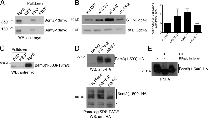
Cdc5/Polo kinase suppresses Cdc42 activation. (A) Lysates from the indicated strains were incubated with GST, GST-PBD, or a control “pincer” mutant PBD* (Elia et al., 2003). Bound GAPs were detected by Western blotting. (B) Log phase wild-type (WT), cdc20-3, cdc5-2, and cdc15-2 cells were shifted to 37°C for 3 h and processed for Cdc42 activation. The example is representative of three experiments. Graph shows means ± SEM. The difference between cdc5-2 and cdc15-2 was statistically significant (P < 0.05 by unpaired two-tailed t test). (C) Lysate from a Bem3(1–500)-13myc cdc15-2 strain (arrested 2.5 h at 35°C) was incubated with purified PBD or PBD* as in A. (D) The indicated strains were arrested as in C; shown is a Western blot to detect altered mobility of Bem3(1–500)-3HA. (bottom) Phos-tag SDS-PAGE was used for increased resolution of phosphorylated bands. Asterisks mark nonspecific bands. (E) Bem3(1–500)-3HA was immunoprecipitated from cdc15-2 cell lysates arrested in telophase as in C and incubated with calf intestinal phosphatase (CIP) and/or phosphatase (PPase) inhibitors. IP, immunoprecipitation; WB, Western blot.

