Figure 3.
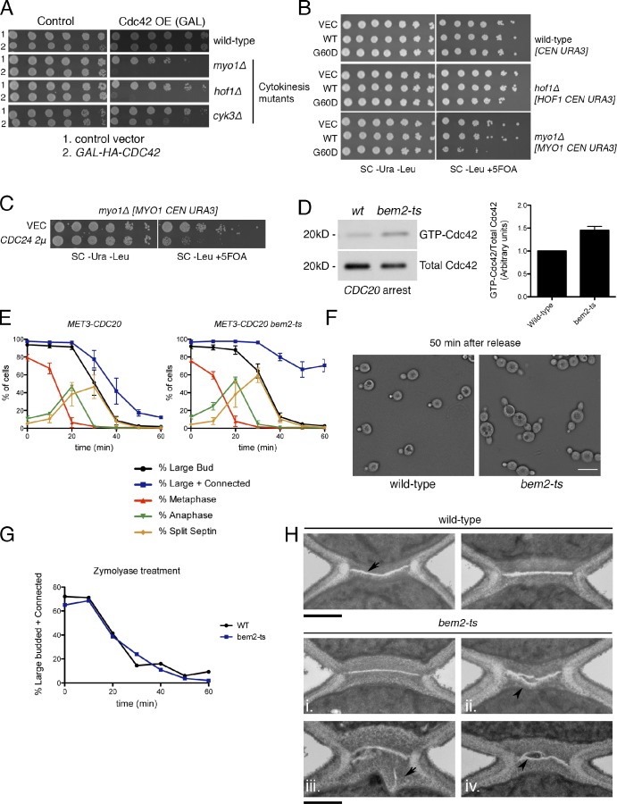
Active Cdc42 interferes with cytokinesis and cell separation. (A) Cells transformed with vector (VEC) or GAL1-HA-CDC42 plasmids were spotted in fivefold dilutions on the indicated media (3 d at 25°C). OE, overexpression; GAL, galactose. (B) Expression of cdc42G60D from the CDC42 promoter on a CEN plasmid (one to three copies per cell) is toxic to myo1Δ cells. Cells were grown on the indicated media (3 d at 25°C). Synthetic lethality is detected on 5-fluoroorotic acid (5FOA) media as a counterselection for the indicated URA3-containing plasmids. (C) myo1Δ cells transformed with control or CDC24 2μ plasmids were grown on plates as in B. SC, synthetic complete. (D) Wild-type (wt) and bem2-ts cells were arrested in metaphase by CDC20 depletion, shifted to the restrictive temperature (37°C at 1.5 h), and processed for Cdc42 activation. Graph shows means ± SEM from three experiments. (E) Cells were arrested in metaphase, shifted to 37°C for 1 h, and released at 37°C. The percentage of cells with the indicated morphology is plotted as means ± SEM from three experiments. Connected indicates two adjacent cell bodies showing evidence of rebudding or repolarization. (F) Bright-field image of cells 50 min after release. Bar, 15 µm. (G) Cells from E were treated with zymolyase to digest the cell wall, and the percentage of large-budded or connected cells was scored. (H) Wild-type cells have a PS (arrow) sandwiched by secondary septa; bem2-ts cells have misaligned, multiple, or otherwise abnormal PSs (arrowheads). Bars, 500 nm.

