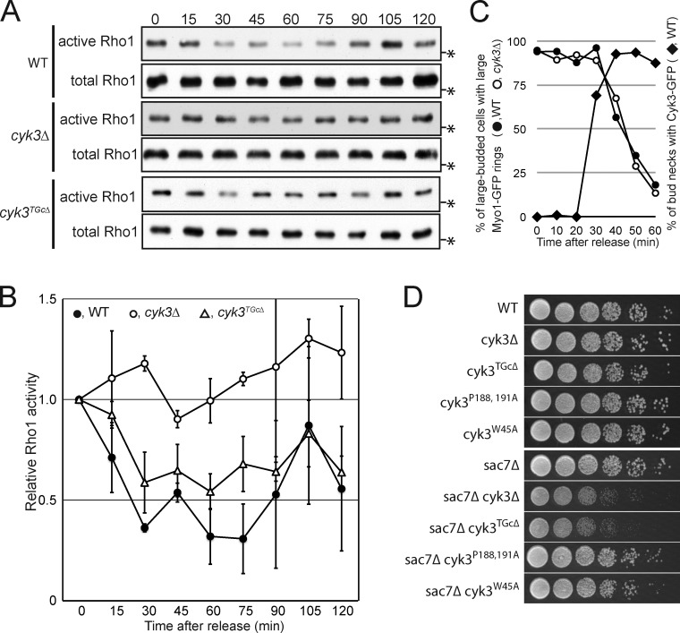
Figure 6.
Temporary inactivation of Rho1 during PS formation and its apparent regulation by Cyk3. (A and B) Activity of Rho1 during cytokinesis in wild-type, cyk3Δ, and cyk3TGcΔ cells. cdc15-2 3HA-RHO1 (MOY553), cdc15-2 3HA-RHO1 cyk3Δ (MOY552), and cdc15-2 3HA-RHO1 cyk3TGcΔ (MOY973) strains were grown to exponential phase in YM-P medium at 24°C, arrested by incubation at 37°C for 3 h, and then released into mitotic exit by cooling rapidly (∼5 min) to 24°C. Samples were collected at the indicated times (minutes) and subjected to the GST-RBD pull-down assay (see Materials and methods). Numbers of independent experiments: WT, 3; cyk3Δ, 2; and cyk3TGcΔ, 2. (A) Representative Western blots of active and total Rho1. Asterisks show the position of a 25.9-kD molecular mass marker. (B) Quantification of band intensities (means ± SDs) of active Rho1 relative to total Rho1; the values at time 0 were set to 1.0. (C) Normal initiation of Myo1-GFP constriction in cyk3Δ cells and timing of Cyk3 localization to the bud neck. cdc15-2 MYO1-GFP (MOY720), cdc15-2 MYO1-GFP cyk3Δ (MOY721), and cdc15-2 CYK3-GFP (MOY543) strains were synchronized as in A. For Myo1-GFP, large-budded cells were scored for the presence of large (i.e., precontraction) or smaller GFP rings (n = 64–111 per sample). For Cyk3-GFP, large-budded cells were scored for the presence of detectable GFP signal at the neck (n = 124). (D) Genetic interaction between CYK3 and SAC7. Strains of the indicated genotypes were spotted on YPD plates as in Fig. 2 A and incubated at 24°C for 48 h (strains: YEF473B, MOY585, MOY882, MWY636, MWY1412, MOY405, MOY440, MOY967, MOY980, and MOY982). WT, wild type.

