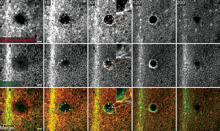
Figure 1.
Drosophila pupal wound healing. Movie stills of a wounded pupal notum expressing mCherry-Moesin under the control of pnr-GAL4 and Sqh-GFP show that pupal epithelial repair recapitulates embryonic wound healing. White arrowheads highlight the actomyosin cable. Actin-rich protrusions, such as lamellipodia and filopodia, are highlighted in the zoom panel at 30 min after wounding. Time after wounding is indicated in the top panels. Bars: (main panels) 20 µm; (insets) 10 µm.

