Figure 8.
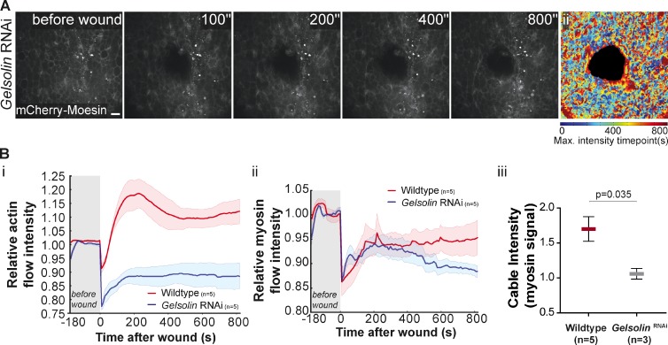
Gelsolin links calcium to the cytoskeleton remodeling. (A) Movie stills of a wounded pupal notum expressing dsRNA against Gelsolin and mCherry-Moesin driven by pnr-GAL4. The actin filament polymerization is not detected as in the control response (Fig. 4 A), indicating that the actin flow is disrupted by Gelsolin down-regulation. (Aii) An impairment of the actin flow can be seen on the graphic representation, color coded as Fig. 2 Aii. Bar, 10 µm. (Bi and Bii) Graphs representing the variation of actin and myosin flow intensity in WT and Gelsolin RNAi showing that upon Gelsolin down-regulation both flows are affected. Shadows represent the SEM for each curve. (Biii) Quantification of myosin cable intensity in WT and Gelsolin RNAi shows that this structure is weaker when Gelsolin expression is reduced (P = 0.035, Mann-Whitney test).

