Figure 1.
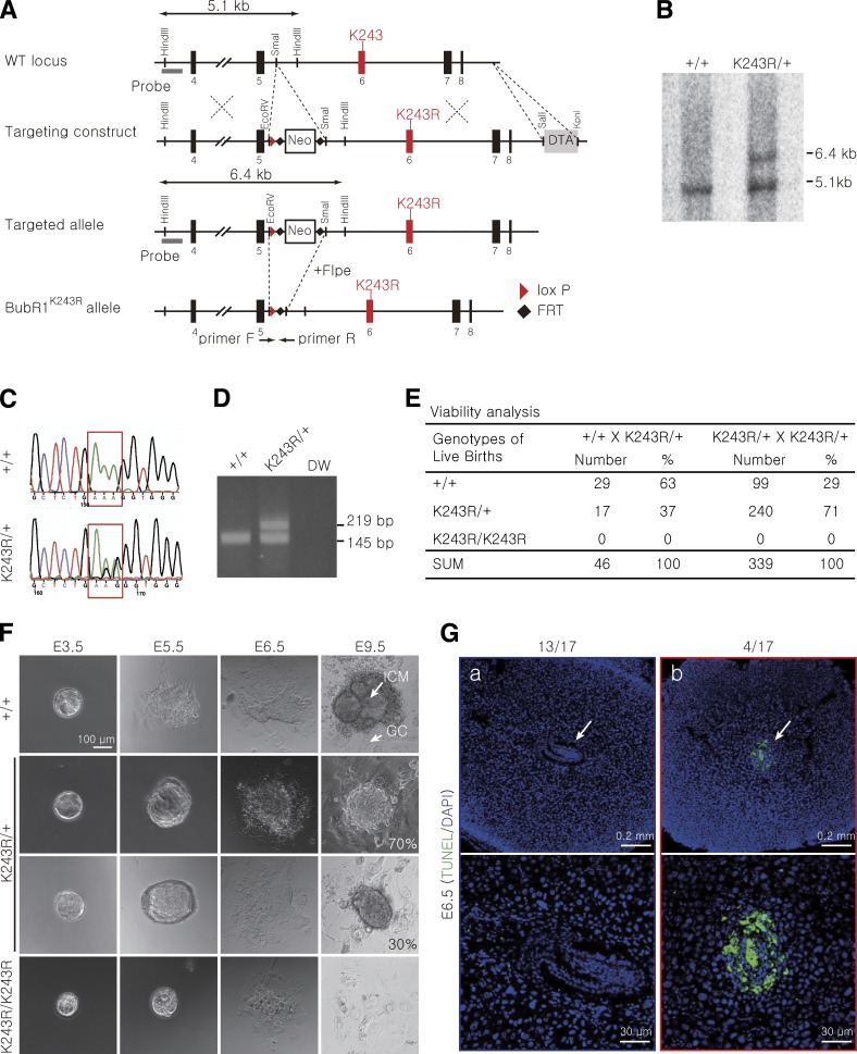
BubR1 acetylation is essential for embryonic development. (A) Schematic representation of the BubR1 gene-targeting strategy. Shown are the structures of the WT BubR1 locus, the targeting construct, the targeted locus, and the disrupted locus after the Flpe-mediated deletion. The neomycin-resistance gene in the targeting construct is removed when crossed with the Flpe transgenic mouse to generate the BubR1K243R (K243R) allele. The probe for Southern blot analysis and the primers for PCR genotyping are marked with gray bars and arrows, respectively. (B) Southern blot analysis of the WT and targeted ES cell lines using the probe indicated in A. HindIII-digested genomic DNA generated a 5.1-kb fragment in the WT and a 6.4-kb band in the targeted allele before the removal of the neo-resistance cassette. (C) Sequence analysis of genomic DNA from WT and K243R/+ ES cells. (D) Genotyping of WT and K243R/+ mice using PCR. (E) Summary of the crosses and progeny. The K243R/+ heterozygous mice were intercrossed, and the newborn pups were scored. (F) In vitro culture of the WT and K243R/+ embryos. The E3.5 embryos from the K243R/+ intercrosses were cultured in vitro. The inner cell mass (ICM) and the trophoblast giant cells (GC) are marked. (G) Sagittal sections of the E6.5 embryos in the uterus were examined for apoptosis using TUNEL staining. Low (top) and high (bottom) power magnification images are shown. The arrows indicate embryos or embryonic remnants.

