Figure 1.
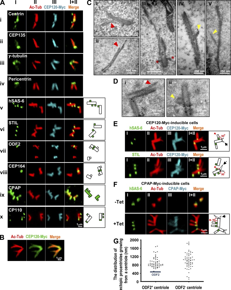
Overexpression of either CEP120 or CPAP induces atypical centriole amplification with extra long centrioles. (A–E) CEP120-Myc–inducible cells were treated with Tet for 48 h and analyzed by confocal fluorescence microscopy using the indicated antibodies (A, B, and E) or by EM (C and D). (C and D) The branched or vertically aligned MT-based filaments are marked by red or yellow arrowheads, respectively. Red asterisks in C (iii) represent appendage structures. (D) The small black dots are nonspecific precipitates that possibly formed during sample preparation. (C) The mean length and width of these CEP120-induced filaments were ∼1,000 and ∼195 nm, respectively (n = 40). (F) Excess CPAP induces the formation of atypical supernumerary centrioles from a preexisting centriole. CPAP-Myc–inducible cells were treated without (−Tet) or with (+Tet) Tet for 48 h and analyzed by confocal fluorescence microscopy. (G) The distribution of atypical ectopic procentrioles growing from ODF2-positive or -negative centrioles (n = 119 from a single experiment). CEP120-Myc–inducible cells were treated with Tet for 48 h and immunostained with antibodies against ODF2 and Ac-Tub.

