Fig. 2.
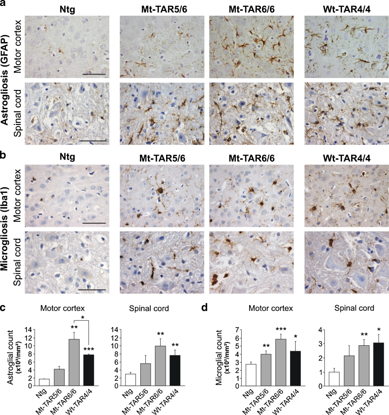
Dose-dependent gliosis in mutant TDP-43 mice. a GFAP immunohistochemistry of cortical layer V and spinal cord of Ntg, Mt-TAR6/6, Mt-TAR5/6, and Wt-TAR4/4 mice showing TDP-43 dose-dependent astrogliosis. b Similarly, Iba1 immunoreactivity showed a dose-dependent microgliosis in Mt-TAR6/6, Mt-TAR5/6, and Wt-TAR4/4 mice compared to Ntg littermates. c Highly increased number of astrocytes in brain and spinal cord of Mt-TAR6/6 and Wt-TAR4/4 mice compared to Ntg littermates. d Significantly increased number of activated microglia in brain and spinal cord of Mt-TAR6/6 and WT-TAR4/4 mice compared to Ntg littermates. Overall, mutant TDP-43 induced a more pronounced gliosis than wild-type TDP-43 overexpression. Data are represented as mean ± SD. Scale bars 50 μm. *P < 0.05; **P < 0.01; ***P < 0.001

