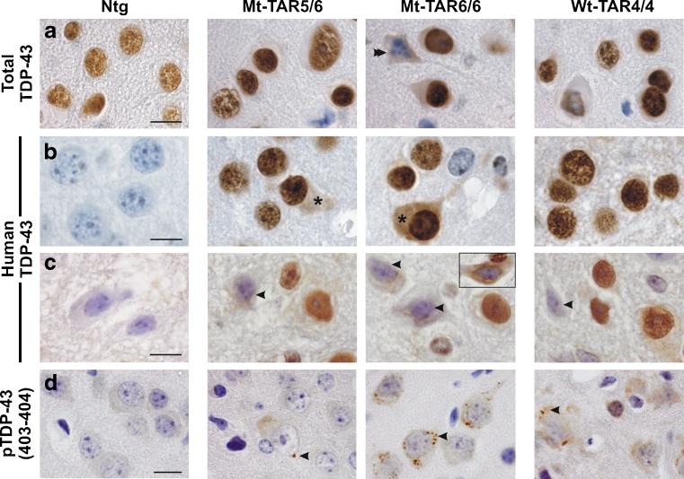
Fig. 4.
Redistribution of nuclear TDP-43 and formation of phosphorylated (pS403/404) TDP-43 accumulations in the cortex of mutant TDP-43 mice. Immunohistochemistry for a total and b–c human TDP-43 of Mt-TAR6/6, Mt-TAR5/6, and Wt-TAR4/4 mice indicated that TDP-43 resides mainly in the nucleus of a–b cortical and c spinal motor neurons. However, nuclear TDP-43 was absent in a apoptotic neurons (double arrowhead). b Using a human-specific TDP-43 antibody, some neurons of Mt-TAR6/6 and Mt-TAR5/6 mice also showed diffuse reactivity in the cytoplasm (asterisks). Human TDP-43 reactivity was absent in Ntg control mice. c The presence of cytoplasmic TDP-43 staining was often accompanied by reduced nuclear TDP-43 immunoreactivity in spinal motor neurons (arrowheads). Nuclear clearing and diffuse cytoplasmic staining were also observed using a non-species-specific antibody (inset). d Small granular structures immunoreactive for phosphorylated (pS403/404) TDP-43 were present within the cytoplasm of cortical neurons of Mt-TAR6/6 and Wt-TAR4/4 mice and, to a lesser extent, in Mt-TAR5/6 mice (arrowheads). Scale bars 10 μm

