
Figure 3.
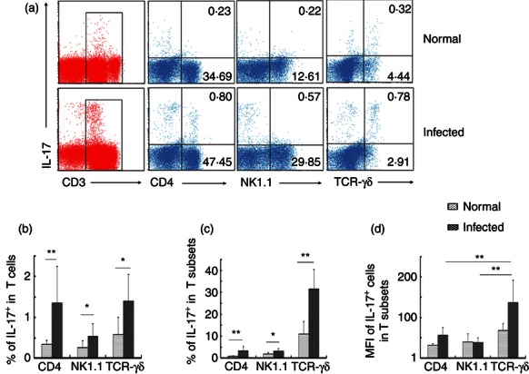
Interleukin-17 (IL-17) expression in specific T-cell subsets isolated from control or infected mouse liver. Female C57BL/6 mice were infected with 40 ± 5 Schistosoma japonicum cercariae per mouse. Six weeks after the infection, the mice were killed. Single-cell suspensions of liver cells were stimulated with PMA, ionomycin and brefeldin A. Cells were stained with anti-CD3-allophycocyanin-Cy7, anti-T-cell receptor-γδ-FITC, anti-CD4-Peridinin chlorophyll protein-Cy5.5, anti-NK1.1-phycoerythrin-Cy7 and then intracellularly stained with anti-IL-17A-phycoerythrin for FACS analysis. (a) Intracellular IL-17 expression by gated populations of CD4+ T cells, natural killer T (NKT) cells and γδT cells isolated from normal and infected mice, respectively. The data are representative of eight experiments giving similar results. (b) Percentage of the IL-17+ NK1.1+, IL-17+ γδTCR+, IL-17+ CD4+ cells in CD3+ T cells were calculated from eight independent experiments with similar results. (c) The percentage of IL-17+ cells in CD4+ T cells, NKT cells and γδT cells were shown. (d) The percentage of mean fluorescence intensity of IL-17+ cells in CD4+ T cells, NKT cells and γδT cells (*P < 0·05, **P < 0·01, the error bars indicate SD).