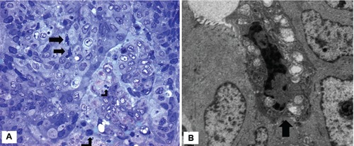
Figure 2.

A) Single adenocarcinoma cells show convoluted nuclei and cytoplasmic vacuolization (arrows). A few neutrophils are seen in the tumour stroma (case 3). Semithin section; Giemsa x400; B) single adenocarcinoma cell characterized by marked chromatin condensation, convoluted nucleus, loss of microvilli and mitochondrial swelling (arrow). The adjacent tumor cells appear morphologically well preserved (case 4), x6000.
