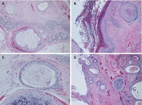
Figure 3.

Histological photographs of the tumor. Hematoxylin and Eosin (H&E) stains at low power (40x, A, D) show lymphoid tissue with metastatic germ cell tumor composed of mixture of respiratory, and gastrointestinal type epithelium, smooth muscle, and cartilage. Higher magnification (H&E, 100x) of the tumor showing squamous, and gastrointestinal type epithelium, and cartilage (B, C).
