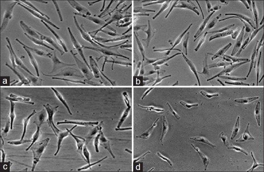
Fig. 3.

Morphological changes of normal human melanocytes HEMa- LP exposed to different kanamycin concentrations for 24 h.
Cell monolayer treated with the lowest concentration of kanamycin (b - 0.06 mmol/l) is similar to the control culture (a - free of antibiotic). The higher concentrations of kanamycin induced detectable changes in cell morphology (c - 0.6 mmol/l, d - 6.0 mmol/l). Cells were observed under an inverted microscope at ×200 magnification.
