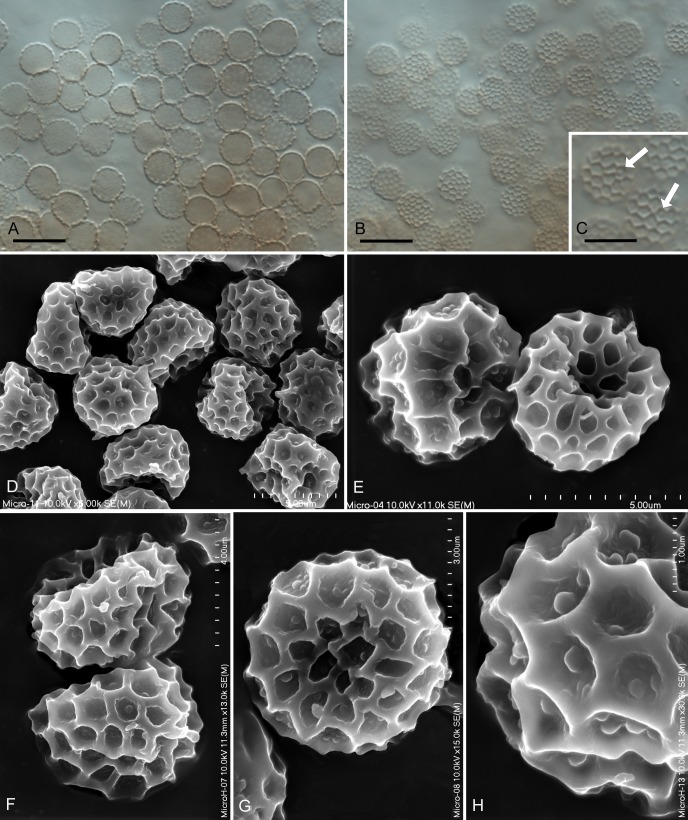
Fig. 3.
Microbotryum silenes-saxifragae sp. nov. on Silene saxifraga (KR-M-23890 – holotype). A–B. Spores seen by LM, median and superficial views. C. Hardly visible tubercles in LM at very high magnification using Nomarski optics, indicated by arrows. D–G. Spores with tuberculate, rough and verruculose interspaces seen by SEM. H. Close-up of spore ornamentation seen by SEM. Bars: A–B = 10 μm, C–E = 5 μm, F = 4 μm, G = 3 μm, H = 1 μm.

