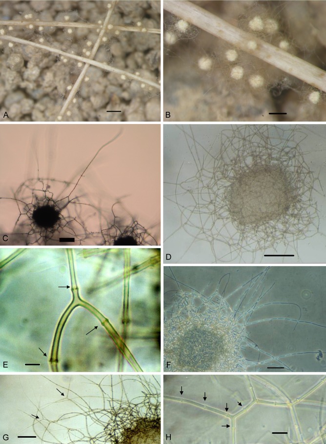
Fig. 1.
Auxarthronopsis bandhavgarhensis (AMH 9405). A–B. Stereomicroscopic-view of mature ascoma growing on horse hair. C. Light microscopic view of unmounted ascomata with elongate appendages picked up from hair bait. D. Mesh-like reticuloperidium with central ascospore mass. E. Base of elongate appendage showing inverted Y-shaped arch with swollen septa (arrows). F. Phase contrast image of ascoma showing multiseptate peridial appendages. G. Bifurcate branching of perdial appendages (arrows). H. Dichotomously branched perdial hyphae showing knuckle joints (arrows). Bars: A = 600 μm; B = 200 μm; C = 100 μm; D = 80 μm; E = 6 μm; F = 80 μm; G = 80 μm; H = 10 μm.

