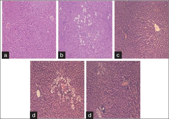
Figure 1.

Hematoxylin and eosin staining, X40. (a) Normal control, normal histological findings in the liver parenchyma. (b) CCl4, submassive confluent necrosis and mixed inflammatory infiltration. (c) Legalon+CCl4, perivenular microvesicular steatosis without necrosis. (d) Hepatisan+CCl4, ballooning inflammatory infiltration and lytic necrosis. (e) Boldo tea+CCl4, ballooning degeneration, inflammatory infiltration, and lytic necrosis
