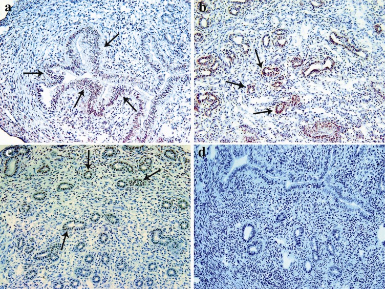
Figure 1.
Immunohistochemical detection of IDO-positive cells in the endometrium of pregnant mice at different gestational periods. Uteri of syngeneic pregnant (Balb/c× Balb/c) mice were removed at early (a), mid (b) and late (c) gestational periods and immunostaining for IDO was carried out on the cryosections. In negative control slides (d), primary antibody was pre-adsorbed by an immunizing peptide with a 50-molar concentration. Black arrows show IDO positive cells (Magnification: 200 ×).

