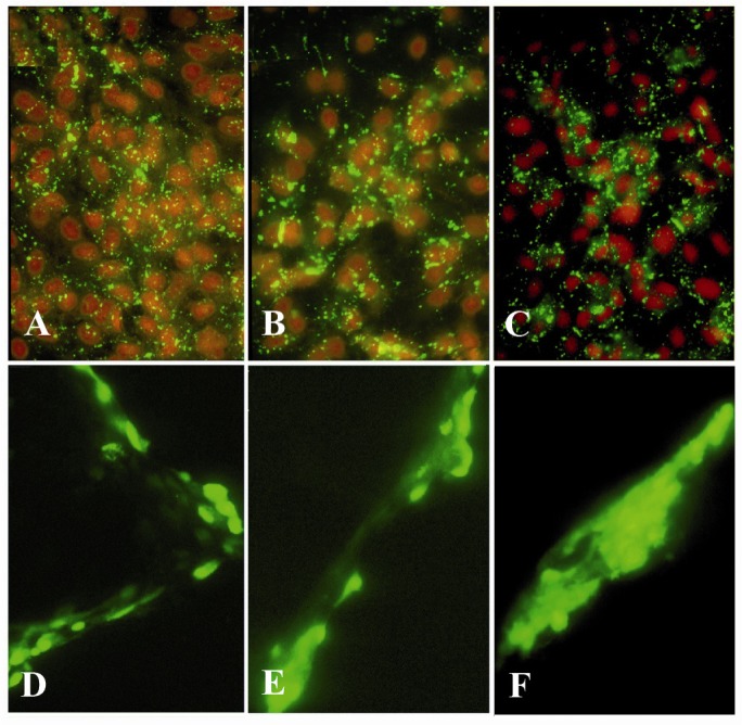
Figure 2.
Immunolocalization of Cx43 (A-C) and lucifer yellow dye transfer (D-F) in GCs on day 4 of culture. GCs were plated on EHS-drip without androstenedione (A & D), with 10−7 M (B & E) or 10−5 M (C & F) androstenedione. Although Cx43 distribution was not altered in either of the cultured conditions, GJIC was markedly enhanced when cells were treated with 10−5 M (F) androstenedione. Fluorescent microscopy images were obtained at 10X (A-C) and 20X magnifications (D-F)

