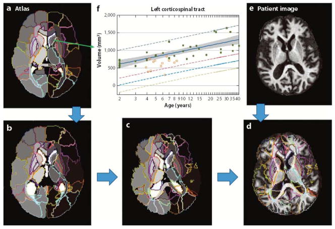
Figure 3.
Whole-brain parcellation based on a preparcellated atlas. An atlas of choice (a), which defines the parcellation criteria, is elastically Warped to an individual image (e) and automatically parcels the entire brain into various structures. The images shown in panels b through d describe the transformation steps for automated parcellation. As the atlas is applied to many clinically normal and abnormal cases, the quantification results, such as the structural volumes of each parcel, are stored in the atlas, from which the average and the standard deviation of normal cases at each age can be characterized (f) (104).

