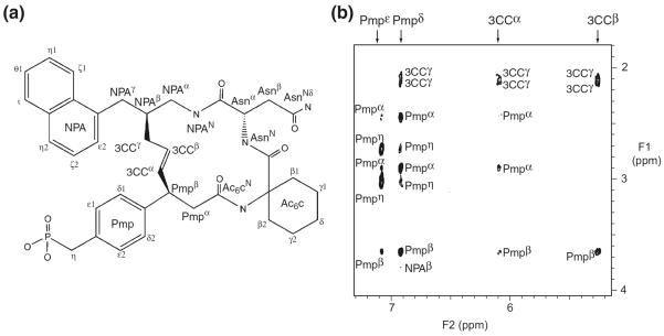
Fig. 3.
(a) Chemical structure of the inhibitor in which the protons were labeled. All proton signals were assigned by NMR. In the inhibitor used for the X-ray study, the phosphonomethyl group was replaced with a malonyl group and α-CH2CO2H was attached to the pTyr-mimicking portion. (b) Expanded view of 2D NOESY spectrum recorded using the complex of the inhibitor and 2H/15N-labeled Grb2 SH2 domain in a 2H2O solution. Inter- and intra-residue NOE connectivity of the inhibitor was labeled

