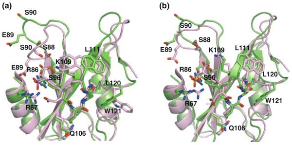
Fig. 7.

Superposition of the three dimensional structures of Grb2 SH2 domain with the inhibitor solved by NMR (green) and X-ray (pink, PDB ID 2AOB). Subunit-A and subunit-B of the crystal structures were shown in panel (a) and (b), respectively. Residues involving in recognition of the inhibitor were shown in stick models
