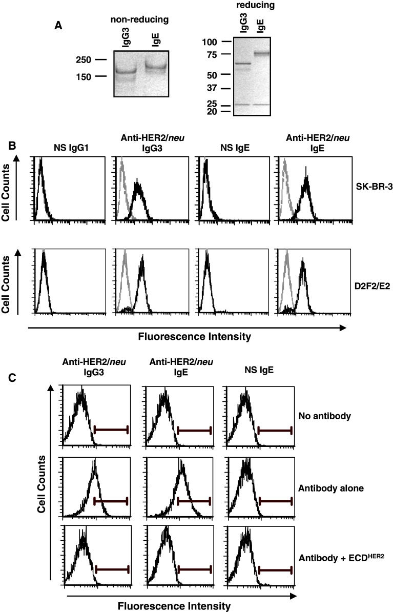
Fig. 1.
Binding to HER2/neu on the surface of cancer cells. a SDS–PAGE analysis of the anti-HER2/neu IgE under both reducing and non-reducing conditions as compared to the anti-HER2/neu IgG3. b SK-BR-3 (top panel) and D2F2/E2 (bottom panel) cells were incubated with 1 μg of a non-specific (NS) IgG1, anti-HER2/neu IgG3, anti-HER2/neu IgE, or a NS IgE. Binding was detected using an anti-human κ-FITC antibody. Samples were analyzed by flow cytometry. Each histogram shows cells incubated with labelled secondary antibody alone (gray) and experimental antibody (black). Data are representative of three independent experiments. c D2F2/E2 cells were incubated with 0.1 μg of the anti-HER2/neu IgG3, anti-HER2/neu IgE, or a NS IgE with or without tenfold excess (1 μg) soluble ECDHER2. Binding was detected using an anti-human κ-FITC antibody. Samples were analyzed by flow cytometry. Data are representative of three independent experiments

