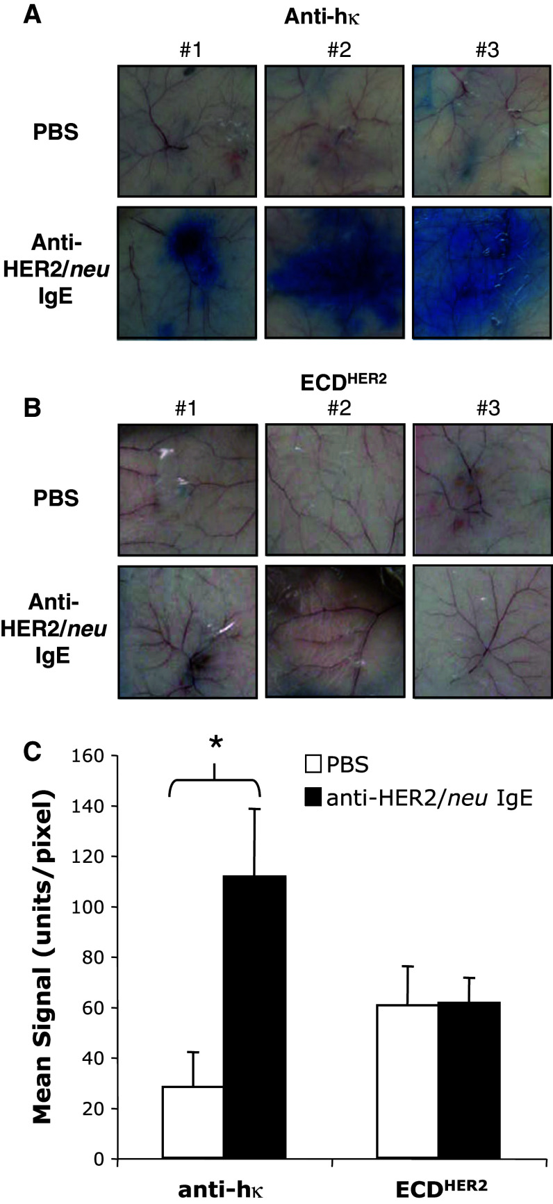
Fig. 4.
IgE-mediated passive cutaneous anaphylaxis in human FcεRIα transgenic animals. Images of the skin of from mice injected intradermically with PBS or 1 μg anti-HER2/neu IgE in a volume of 50 μl. Panels 1, 2, and 3 show images from three different animals per treatment. After 4 h, a 25 μg of anti-human kappa (hκ) or b 2 μg ECDHER2 was injected in 1% Evan’s Blue in PBS. The mice were euthanized after 10 min. c Leakage of the blue dye into the skin due to a local IgE-induced inflammatory response was assessed using the NIH ImageJ Software (graph corresponds to the images shown). The color intensity is reported as the mean signal per pixel and is the average of three different animals corresponding to the images shown in (A) and (B). *P < 0.01 Student's t test

