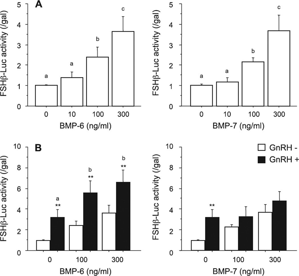
Fig. 1.
Effects of BMPs and GnRH on FSH-induced transcriptional activity in LβT2 cells. After preculture, cells (1.5 × 10⁵ viable cells/cm2) were transiently transfected with 500 ng of FSHβ-Luc and 50 ng of cytomegalovirus-β-galactosidase plasmid (pCMV-β-gal). The cells were then treated with indicated concentrations of BMP-6 and BMP-7 in the (A) absence or (B) presence of GnRH (10 nM) for 24 h. Cells were washed with PBS and lysed, and the luciferase activity and β-galactosidase (β-gal) activity were measured by a luminometer. Results are shown as the ratio of luciferase to β-gal activity and graphed as means ± SEM of data from at least three separate experiments, each performed with triplicate samples. For each result within panels (A) and (B), values with different superscript letters are significantly different at P ≤ 0.05, and for each result within panel B), **P ≤ 0.01 vs. GnRH (−) group.

