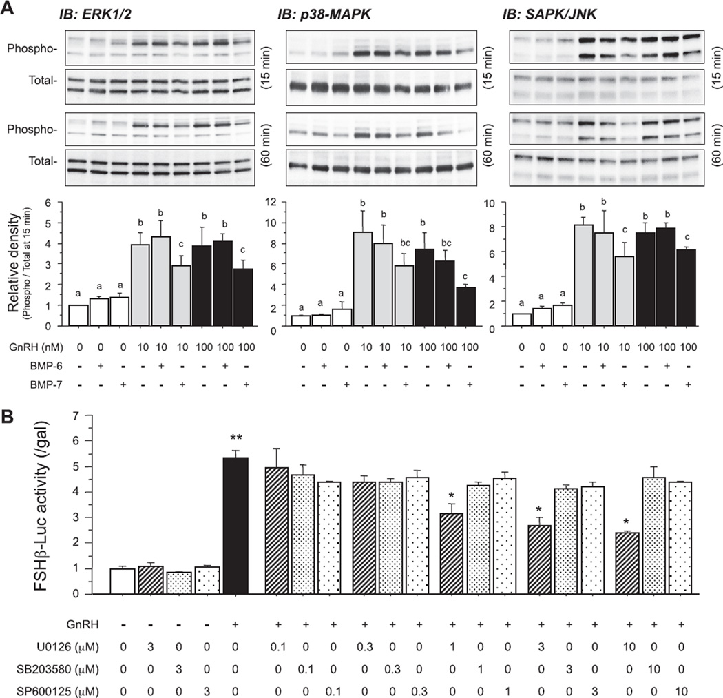
Fig. 3.
Effects of BMPs on GnRH-induced MAPK pathways in LβT2 cells. (A) Cells (3 × 10⁵ viable cells/cm2) were precultured for 24 h and treated with BMP-6 and BMP-7 (100 ng/ml) in the absence or presence of GnRH (10 and 100 nM). After 15- and 60-min culture, cells were lysed and subjected to SDS–PAGE/immunoblotting (IB) analysis using anti-phospho-ERK1/2 and anti-total-ERK1/2, anti-phospho-p38 and anti-total-p38, and anti-phospho-SAPK/JNK and anti-total-SAPK/JNK antibodies. The results shown are representative of those obtained from three independent experiments. The relative integrated density of each protein band at 15-min stimulation was digitized by NIH image J 1.34s and shown as phospho-/total-protein levels in each panel. Results are shown as means ± SEM of data from at least three separate experiments, each performed with triplicate samples. For each result within panel (A), the values with different superscript letters are significantly different at P < 0.05. B) After preculture, cells (1.5 × 10⁵ viable cells/cm2) were transiently transfected with 500 ng of FSHβ-Luc and 50 ng of cytomegalovirus-β-galactosidase plasmid (pCMV-β-gal). The cells were then treated with indicated concentrations (µM) of MAPK inhibitors including U0126, SB203580 and SP600125 in the presence of GnRH (10 nM) for 24 h. The cells were washed with PBS and lysed, and the luciferase activity and β-galactosidase (β-gal) activity were measured by a luminometer. Results are shown as the ratio of luciferase to β-gal activity and graphed as means ± SEM of data from at least three separate experiments, each performed with triplicate samples; *P < 0.05 and **P < 0.01 in each set of the bar graphs within panel (B).

