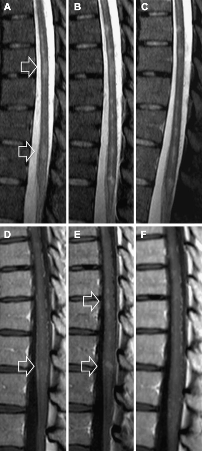
Figure. MRI of the lower spinal cord.

Sagittal STIR (A–C) and T1-weighted sequences after gadolinium (D–F) at the acute stage (A and D), at day 15 (B and E), and at 2 months (C and F). The initial MRI (A, D) shows dorsal hyperintensities (A: empty arrows) with slight enhancement of the lower one (D: empty arrow), leading to a diagnosis of multiple sclerosis relapse. The second MRI (after administration of methylprednisolone) (B, E) shows increased size of previous hyperintensities and new hyperintensities in STIR (B) and new gadolinium enhancements in T1-weighted sequences (E: empty arrows). At this time, the diagnosis of varicella-zoster virus myelitis was made and acyclovir treatment was initiated. The final MRI (after acyclovir/valacyclovir treatment) shows decreased size of hyperintensities in STIR (C) and a marked decrease of contrast enhancements (F).
